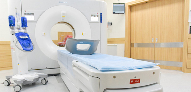
CT
-
LocationUnit1 (1F of the main building), Unit2 (1F of the east building & main building)
-
PurchaseApril, 2020
Overview
Spectral CT scanner(the IQon Elite Spectral CT) provides a more nuanced look at body tissues, which reveals some information about the composition of anatomy and not only its structure. The different frequencies of X-rays in a spectral CT can be compared to different colors of light, which are produced by different frequencies of visible radiation. In a spectral CT, things like iodine show up at lower energies, while the visual impact of metal can be reduced when viewing the higher energy data.
Features
Diagnostic certainty
With IQon Elite Spectral CT, true conventional CT images are paired with layers of spectral results in the same scan. Suddenly, what had been invisible is now both visible and diagnostic. IQon Elite Spectral CT’s improved tissue characterization and visualization provides diagnostic certainty far beyond that of conventional CT.
Every scan is a spectral scan
Only IQon Elite Spectral CT eliminates the need for upfront clinical decision-making through the revolutionary NanoPanel Prism detector. With detector-based spectral technology, only IQon Elite Spectral CT solves the pre-scan dilemma—delivering the certainty of true spectral data in every single scan.
Powerful advancements that fit your workflow
With no special training or separate scanning modes required, IQon Elite Spectral CT integrates seamlessly into your current workflow practice to help you establish a new, spectral standard of care. Utilizing tools that enable on-demand, simultaneous viewing and quick comparison of multiple spectral results from a single region of interest, clinicians have easy access to rich data that can improve confidence in findings.
HyperSight Elite Spectral Reconstructor
Designed to address the needs of high throughput and emergency care settings, our the IQon Elite Spectral CT's Elite Spectral Reconstructor enables routine spectral imaging of up to 200 patients in a 16-hour shift.
Calcium Suppression
This spectral result provides images that provide additional information to the clinician that may help in better assessment of intervertebral disc herniation, and allows for better visualization of bone marrow pathology.
Electron Density
This spectral result provides images that are created using a dedicated algorithm to estimate the electron density of each voxel, helping to enhance tissue characterization and enabling a new level of diagnostic certainty for oncology clinicians and their patients.
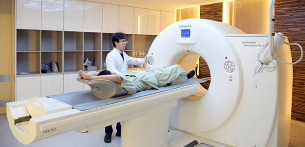
-
LocationUnit1 (2F of the west building)
-
PurchaseDecember, 2015
Overview
CT Scanner provides fast and accurate scanning with dose reduction, maximizes patient safety The Siemens SOMATOM Definition Flash is a whole-body CT scanner that optimizes scan time and dose efficiency, allowing for successful imaging on a variety of patients. With thorax scans that take approximately 0.6 seconds, clinicians can get clear images, without a breath hold if necessary — increasing the scanner’s performance possibilities in trauma and critical care settings.
Features
Redefining standards in Cardiology
When performing a cardiac scan, the main challenge is often posed by the time and effort it takes to prepare the patient. With a scan time of only 0,6 s and a full temporal resolution of 75 ms, the new SOMATOM Definition Flash allows scanning the entire heart and evaluating its morphology and coronaries without any beta blockers. Patients suffering from irregular heart rates or atrial fibrillations benefit from high-end cardiac scanning only taking 250 ms to perform – about a quarter of a heartbeat. Redefining your standards in cardiology with the new SOMATOM Definition Flash will expand your institution’s clinical capabilities, increase outcome quality for your patients, and help you streamline workflows significantly.
Brian B. Ghoshhajra, M.D.
MGH Boston, Massachusetts, USA
Redefining standards in Pediatrics
A key challenge in pediatrics is to get these young patients to hold still. That’s why today’s standard procedure includes intense preparation, sedation, and after-care. The Flash Spiral scan mode combines a scan speed 458mm/s and a temporal resolution of 75 ms – reducing the average examination time to only 0.49 s, in effect allowing you to routinely perform pediatric scans without hazardous sedation. Flash speed and FAST CARE technology make pediatric imaging both easier to use and more patient-friendly – for example, FAST 3D Align automatically optimizes the field of view. Increase your institution’s competitiveness and improve patient outcome by making Flash speed and Dual Source your standard.
Marilyn J. Siegel, MD
Pediatric Radiologist, Washington University, USA
Redefining standards in Oncology
In order to provide precise diagnoses, oncology examination is all about recognizing lesions, and visualizing and quantifying tissue. With Dual Source Dual Energy scanning – the only modality that enables DE scanning at doses comparable to a conventional 120 kV scan –, you will be able to get all the information you need in a single scan. Creating virtual non-contrast images based on the Dual Energy information will help you and your institution save time and resources. Dual Energy Optimum Contrast achieves images with very low noise at high contrasts. Enhance your institution’s clinicial capabilities by standardizing dose-neutral Dual Source Dual Energy CT – introducing „two-in-one“ CT scanning for all your patients, and turning imaging information into imaging intelligence with syngo.via applications.
Hirofumi Kuno, MD
National Cancer Center Hospital East, Chiba, Japan
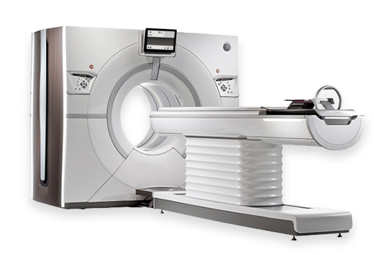
-
LocationUnit1 (1F in the Emergency Center of the east building)
-
PurchaseAugust, 2017
Overview
Get uncompromised image quality and clinical capabilities through the convergence of coverage, spatial resolution, temporal resolution and spectral imaging – all in one system. What if your CT could address the needs of all your patients, even the challenging ones? What if your CT could help you deliver clinical excellence across all of your departments? It’s time for a Revolution. Revolution CT delivers uncompromised image quality and clinical capabilities through the convergence of coverage, spatial resolution, temporal resolution and spectral imaging – all in one. It is the CT designed to help you deliver revolutionary and differentiated capabilities across all of your clinical areas.
Features
GSI Xtream
GSI (Gemstone Spectral Imaging), was introduced on the Discovery 750HD in 2010. This technology allows for fast kV switching during dual-energy data acquisition. This, in turn, allows near-simultaneous dual-energy projections, minimizing the effect of organ and patient movement.
The original GSI from the 750HD has been improved and integrated into the Revolution series as GSI Xtream. Revolution CT scanners are the first to include this technology. Reduction in metal artifacts, improved lesion detection, and .25 millisecond kV switching are a few of the benefits made possible by GSI Xtream.
Smartdose
The integration of ASiR (GE's proprietary iterative reconstruction software) helps to improve low-contrast detectability and reduce dose by more than 80%. The result is shorter exams as low as 70 kV- perfect for fast and safe pediatric imaging.
Smart Patient Centering (SPC)
This feature helps to detect off-center imaging prior to each patient scan. The feature suggests table movements to the tech to correct the centering, allowing Revolution CT users to avoid taking unnecessary scans and exposing patients to unnecessary radiation dose.
Organ Dose Modulation (ODM)
ODM is a capability within the Smartdose feature family. The software adjusts radiation dose according to the presence of certain organs in the path of the scan. This helps reduce overall dose and protects sensitive areas from unnecessary radiation.
Smart Flow
For techs using Revolution series scanners, Smart Flow is a potential game changer. The software improves patient throughput by accommodating a broader scan area up to 200 cm. Smart Flow also includes tabbed workflows to make managing multiple patients simpler.